How to Cite
License

This work is licensed under a Creative Commons Attribution-ShareAlike 4.0 International License.
Identifiers
Published
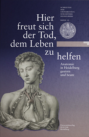
Hier freut sich der Tod, dem Leben zu helfen
Anatomie in Heidelberg gestern und heute
The study of human anatomy, of the relationships and connections between organs, tissues and cells, together with their structures, was established in the 16th century as a fundamental component of medical research and teaching. Since then, anatomy methodologies have changed considerably. Where once it was sufficient to expose components of the body with a dissecting knife and simply describe what was seen with the naked eye, current-day anatomists have access to many additional, highly sophisticated methods for observation and measurement including, for example electron microscopy and computer tomography. These new methods have opened up many new possibilities of analysis and investigation for this area of science.
This exhibition includes many different aspects of anatomy: in addition to the current activities of the Heidelberg Institute for Anatomy and Cell Biology in teaching and research, it covers the Institute’s history going back to the year 1805. The focus of the historical component of the exhibition is on the Institute’s past directors, whose research and publications made many influential contributions to the development of the field. A third component of the exhibition introduces the anatomical preparations and models that have been an important component of applied anatomy since the 18th century. This is presented through a focus on the history and artefacts of The Heidelberg Anatomical Collection. A final component of the exhibition covers the development of anatomical illustrations in printed works ranging from the 16th to 19th century, of which the great majority are the property of Heidelberg University Library.


